
ECG practice: read the strip, then name the rhythm
Take one heartbeat apart on a 3D heart, learn the criteria for 15 rhythms, then test yourself on a new strip every time. Free, in your browser, no account.
P wave
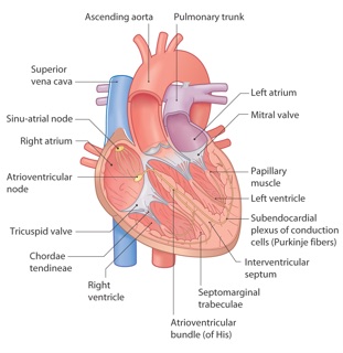
The atria depolarise. The signal starts at the SA node in the right atrium and spreads over both atria.
“This app is so amazing and helps with studying SO SO SO SO much, I wish I could use Lexie for every exam I have so I wouldn't need to stress 24/7”
Shine PemipaGoogle Play Store review
Learn the beat. Learn the rhythms. Then test.
Three parts, in the order a course teaches them
One beat, part by part
Tap the P wave, the PR interval, the QRS complex, the ST segment, the T wave or the QT interval. The 3D heart lights up the chambers that make it, and you read what normal looks like.
Fifteen rhythms with their criteria
Each rhythm comes with its rate, its regularity, its P waves, its PR interval and its QRS, plus the one clue that gives it away. Tap two points on any strip to measure it.
Ten questions, a new strip every time
Name the rhythm from four options, count the rate, name a part of the beat, and tap the right chamber on the heart. Wrong answers show the clue you missed.
How to read an ECG strip in five steps
1. Rate
Count the QRS complexes on a six-second strip and multiply by ten. On a regular rhythm, divide 300 by the number of large squares between two R waves.
2. Rhythm
Check the distance from R wave to R wave along the strip. It is regular, irregular in a pattern, or irregular with no pattern.
3. P waves
Look for one upright P wave before every QRS. No P waves, extra P waves and sawtooth waves each point to a different rhythm.
4. PR interval
Measure from the start of the P wave to the start of the QRS. Normal is 3 to 5 small squares. Check that it stays the same from beat to beat.
5. QRS complex
Measure its width. Under 3 small squares is narrow, and the beat came from above the ventricles. Wider than that, suspect a beat from the ventricles. A bundle branch block also widens the QRS, so check the P waves too.
The 15 rhythms
Normal sinus rhythm
Sinus rhythms. Regular, 60 to 100 per minute, and one P wave before every QRS.
Sinus bradycardia
Sinus rhythms. Every part of the beat is normal. Only the rate is under 60 per minute.
Sinus tachycardia
Sinus rhythms. Rate over 100 per minute, and you can still see a P wave before every QRS.
Atrial fibrillation
Atrial rhythms. No P waves, and no two R-R intervals are the same.
Atrial flutter
Atrial rhythms. A sawtooth baseline, with several flutter waves for each QRS.
Supraventricular tachycardia (AVNRT or AVRT)
Atrial rhythms. Fast, narrow and perfectly regular, with no P waves to see.
First-degree AV block
AV blocks. The PR interval is longer than one large square, and every P wave still gets a QRS.
Second-degree AV block, Mobitz I (Wenckebach)
AV blocks. The PR interval gets longer and longer, then one P wave has no QRS.
Second-degree AV block, Mobitz II
AV blocks. The PR interval never changes, and then a P wave has no QRS, with no warning.
Third-degree (complete) AV block
AV blocks. P waves march through at their own rate, and the slow QRS complexes take no notice of them.
Sinus rhythm with PVCs
Ventricular rhythms. An early, wide QRS with no P wave before it, then a pause.
Ventricular tachycardia (monomorphic)
Ventricular rhythms. Fast, regular and wide.
Ventricular fibrillation
Ventricular rhythms. No P waves, no QRS complexes, no T waves. Only a chaotic line.
Asystole
Ventricular rhythms. A flat or almost flat line. Check the patient, the leads, the gain and a second lead before you call it asystole.
Sinus rhythm with ST elevation
ST segment. The line does not come back to the baseline after the QRS. It stays high and runs into the T wave.
Frequently asked questions
Both mean the same test. ECG comes from electrocardiogram, and EKG from the German spelling. ECG is the usual spelling in most of the world, and EKG is common in the United States. The strips, the waves and the rhythms are the same.
No. Every strip is drawn from a formula with textbook timings, in lead II, at 25 mm per second and 10 mm per millivolt. That keeps each rhythm clean enough to learn from, and it gives you a new strip every time you test. Real recordings have artefact and variation, so practise on those too once you know the criteria.
Count the QRS complexes on a six-second strip and multiply by ten. On a regular rhythm, divide 300 by the number of large squares between two R waves. Each strip here is six seconds long, and you can tap two points to measure the time between them.
Normal sinus rhythm, sinus bradycardia, sinus tachycardia, atrial fibrillation, atrial flutter, supraventricular tachycardia, first-degree AV block, second-degree AV block types Mobitz I and Mobitz II, third-degree AV block, sinus rhythm with PVCs, ventricular tachycardia, ventricular fibrillation, asystole, and sinus rhythm with ST elevation.
Yes, it is free, and you do not need an account. Open the trainer and start. If you want to study your own cardiology notes, drop them into Lexie and it makes a study set from them.
No. This page is for study. It does not diagnose anything, and one lead cannot confirm a diagnosis. For a real patient, get a 12-lead ECG and a clinician.
Lexie wrote the criteria and the explanations with AI, from textbook values, and checked them against standard references. AI can make mistakes. If something here differs from your course material, trust your course material and tell us.
Now study your own cardiology notes
Drop a lecture, a PDF or a photo into Lexie. It picks out the concepts and turns them into flashcards, a quiz and written questions.